

SKIN LINE · CONCERNS
Haemangioma
why it occurs and what we can do about it
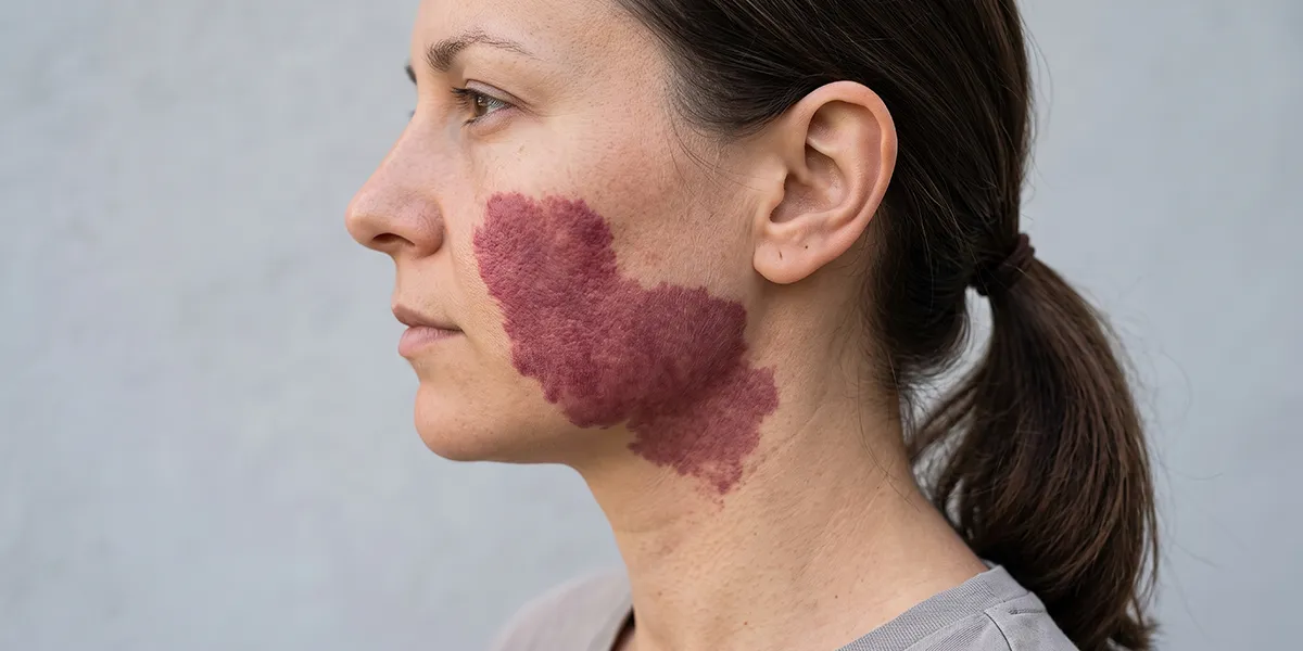
A haemangioma is a benign growth associated with blood vessels that can occur in both children and adults. Depending on the type, it may appear as a small red spot, a clearly defined vascular formation, or a larger growth located in the deeper layers of the skin. Although haemangiomas are not dangerous in most cases, some require monitoring or treatment.
The term ‘haemangioma’ is often used to refer to various vascular growths that are not entirely identical in their origin and behaviour. Therefore, correctly identifying the type and characteristics of the formation is an important first step. Modern dermatology has various methods for monitoring and treating haemangiomas where necessary.
What is a haemangioma?
A haemangioma is a benign vascular growth composed of cells and structures associated with blood vessels. Its appearance depends on which vessels are involved and how deep the growth is located.
Haemangiomas can vary in size and colour. Superficial growths usually have a bright red or purplish hue, whilst those located deeper may appear bluish or be a colour similar to that of the surrounding skin.
When pressure is applied, some vascular lesions may fade slightly, as blood is temporarily forced out of the vessels. However, this is not sufficient to make a diagnosis.
In children, the condition is most commonly referred to as an infantile haemangioma. It may not be visible at birth or may appear during the first few weeks of life. After a period of growth, it usually begins to shrink and fade gradually.
In adults, the red, dot-like lesions often referred to as ‘haemangiomas’ are usually cherry angiomas – common, benign vascular lesions.
Types of haemangioma
Haemangiomas can be classified according to various criteria, including the age of onset, the depth and the characteristics of the vascular formation.
Infantile haemangioma – most commonly appears during the first few weeks of life. It usually goes through a phase of rapid growth, followed by a gradual spontaneous reduction in size. Many of these haemangiomas do not require treatment, but certain locations and sizes do require medical supervision.
Superficial haemangioma – affects the more superficial layers and is usually bright red in colour. In the case of infantile haemangiomas, the term ‘strawberry haemangioma’ is often used to describe such lesions.
Deep haemangioma – is situated deeper within the skin or subcutaneous tissue. It may appear as a bluish or flesh-coloured raised area.
Mixed haemangioma – has both a superficial and a deeper component.
Cherry angioma – a small, benign vascular lesion that is very common in adults. It usually presents as a small red to purple papule and can appear on various parts of the body.
As different vascular lesions can look similar, the diagnosis must be made by a doctor, particularly in the case of a rapidly changing or atypical lesion.
Causes and risk factors for the development of haemangiomas
The exact cause of infantile haemangiomas is not fully understood. They are not considered to be the result of parental behaviour during pregnancy, nor are they caused by skin trauma.
Certain factors have been identified in connection with infantile haemangiomas that are associated with a higher likelihood of their occurrence. These include:
- female sex;
- preterm birth;
- low birth weight;
- certain characteristics of the pregnancy;
- the presence of more than one haemangioma.
It is important to note that the presence of a risk factor does not mean that the child will necessarily develop a haemangioma.
The exact cause of cherry angiomas is also not fully understood. They become more common with advancing age and may occur in several members of the same family.
Hormonal factors and other individual characteristics are likely to play a role in some vascular lesions, but it should not be assumed that every occurrence of a haemangioma is linked to a specific disease or hormonal disorder.
Is a haemangioma a disease?
A haemangioma is usually a benign vascular lesion, not a malignant tumour. In many people, it does not cause any health problems and does not require active treatment.
Infantile haemangiomas follow a characteristic natural course. After the initial growth phase, they usually begin to shrink gradually. The process of involution can take years.
However, not all haemangiomas should simply be left unmonitored. Lesions located near the eyes, nose, mouth or airways may cause functional problems depending on their size and location.
Larger or rapidly growing infantile haemangiomas may ulcerate, bleed or leave permanent changes to the skin. For this reason, early treatment is necessary in certain cases.
When should you see a doctor?
Not every new vascular lesion should automatically be classified as a haemangioma. Consulting a dermatologist is particularly important when the lesion is new, changing in appearance or has an unusual appearance.
See a doctor if:
- the growth is increasing rapidly in size;
- it starts to bleed or develop an ulcer;
- it changes significantly in colour or shape;
- it is painful;
- it is frequently injured by clothing, jewellery or shaving;
- it is located near the eye, nose or mouth;
- causes a functional problem;
- numerous new vascular lesions appear within a short period.
In a baby with a haemangioma, medical assessment is particularly important if the lesion is growing rapidly, is large or is located in an area where it may affect vision, feeding or breathing.
If necessary, the specialist may use a dermatoscope, and in the case of deeper or more complex vascular lesions, further investigations may be recommended.
Daily care at home for haemangiomas
In most cases, a haemangioma does not require any special daily treatment. The most important thing is to protect the area from injury.
If the growth is raised and is located in a place where it can easily catch on clothing or be injured whilst shaving, care must be taken during these activities.
The skin around it can be kept clean and well moisturised, but it is not necessary to use harsh products.
In children with infantile haemangioma, it is important for parents to monitor the size and appearance of the lesion. Taking photographs at regular intervals under consistent conditions can help the doctor track changes over time.
If ulceration or bleeding occurs, do not apply any unauthorised preparations. It is essential to seek medical advice on appropriate care.
Home remedies for managing the condition and home care measures
A haemangioma must not be treated with home remedies intended to ‘burn off’, shrink or remove the growth. Vinegar, acids, essential oils, alcohol and other corrosive substances can cause burns and further damage to the skin.
It is also not advisable to pierce, cut into or apply pressure to the lesion. As it contains blood vessels, trauma to it can lead to bleeding and infection.
The best home care involves protecting the area from injury, maintaining good hygiene and monitoring for any changes. If you suspect an infection, ulceration or bleeding, a medical assessment is required.
In most cases, a haemangioma is a benign condition and does not pose an immediate risk. However, a correct diagnosis is important because various vascular lesions may look similar, and some of them require monitoring or treatment. Modern dermatology offers effective options when removal or medical intervention is necessary. The most appropriate method must always be determined on a case-by-case basis following an examination by a specialist.
Modern solutions for managing haemangiomas
Not every haemangioma needs to be removed. For many, the most appropriate approach is observation, particularly when they are small, cause no symptoms and are not located in a high-risk area.
For infantile haemangiomas that require treatment, modern medicine offers effective pharmacological approaches. In certain cases, propranolol is used; this is the main systemic treatment for problematic infantile haemangiomas. It is prescribed by a doctor following an assessment of the individual case.
For some small and superficial haemangiomas, topical timolol may be used when the specialist deems it appropriate.
Various dermatological procedures may be used to treat certain vascular lesions in adults. These include laser technologies, electrocoagulation and other methods for the controlled removal of superficial vascular lesions. The choice depends on the specific type, size and depth of the lesion.
For some larger or more complex vascular malformations, the approach may differ and may involve specialist treatment by a team with experience in vascular anomalies.
A personalised approach to your skin
Discuss your condition and treatment options with a specialist from Skin Line.
Clinics in Varna, Plovdiv and Sofia.
Treatment options
Your care begins with a consultation
Choose a clinic that suits you and get in touch with our team.

